Right Shoulder Anatomy Diagram : Shoulder Arm Atlas Of Anatomy : This diagram here just shows the joint capsule itself.
byAdmin•
0
Right Shoulder Anatomy Diagram : Shoulder Arm Atlas Of Anatomy : This diagram here just shows the joint capsule itself.. I sustained fractures to the right shoulder & top of arm in 2003. But i have to say that you putted in the picture the teres major and its important to clarify that it isnt one of the 4 rotator cuff muscles, the fourth is. The shoulder joint (glenohumeral joint) is a ball and socket joint between the scapula and the humerus. When you realize all the different ways and positions we use our hands. Shoulder radiology & anatomy at usuhs.mil.
Human anatomical atlas of the shoulder : Normal anatomy, variants and checklist. The shoulder joint is formed where the humerus (upper arm bone) fits into the scapula. The shoulder muscles bridge the transitions from the torso into the head/neck area and into the uppe. The glenohumeral joint has the following supporting structures:
Lateral View Of The Right Shoulder Joint Diagram Quizlet from o.quizlet.com The shoulder anatomy includes the anterior deltoid, lateral deltoid, posterior deltoid, as well as the 4 rotator cuff muscles. Anatomical diagram of the muscles of the neck. The shoulder joint is formed where the humerus (upper arm bone) fits into the scapula. Explore more like shoulder joint anatomy diagram. Ap x ray of a dislocated right elbow. Enjoy the videos and music you love, upload original content, and share it all with friends, family, and the world on youtube. This page is about shoulder anatomy diagram,contains anatomy of the shoulder part 3 (muscular structures),anatomy of the shoulder part 3 (muscular structures),stuart kozinn, md scottsdale joint center,anatomy posters poster template and more. Blank head and neck muscles diagram | body muscles … from i.pinimg.com.
Right shoulder joint arthrography coronal t1wi a coronal t2wi b download scientific diagram the diaphragm and liver in context.
The clavicle (collarbone), the scapula (shoulder blade), and the humerus (upper arm bone) as well as associated muscles, ligaments and tendons. In this episode of eorthopodtv, orthopaedic surgeon randale c. Normal anatomy, variants and checklist. I sustained fractures to the right shoulder & top of arm in 2003. Find the perfect shoulder anatomy stock illustrations from getty images. Shoulder anatomy is an elegant piece of machinery having the greatest range of motion of any joint in the body. Arteriography (angiography) of the right. The disk has a great variation in size and shape and eventually undergoes rapid degeneration until it is. In human anatomy, the shoulder joint comprises the part of the body where the humerus attaches to the scapula.1 the shoulder is the group of structures in the region of the joint.2. We're looking laterally now at the right shoulders. Use the mouse scroll wheel to move the images up and down alternatively use the tiny arrows (>>) on both side of the image to move the images. This acts as the bony framework by which the muscles of the chest, upper back and shoulder connect the upper limb to the trunk of the body and control it's movements.the clavicle connects to the sternum via the. Shoulder radiology & anatomy at usuhs.mil.
Webmd's shoulder anatomy page provides an image of the parts of the shoulder and describes its function, shoulder problems, and more. Wiring diagram for genie garage door opener. We're looking laterally now at the right shoulders. This acts as the bony framework by which the muscles of the chest, upper back and shoulder connect the upper limb to the trunk of the body and control it's movements.the clavicle connects to the sternum via the. Learn vocabulary, terms and more with flashcards, games and other study tools.
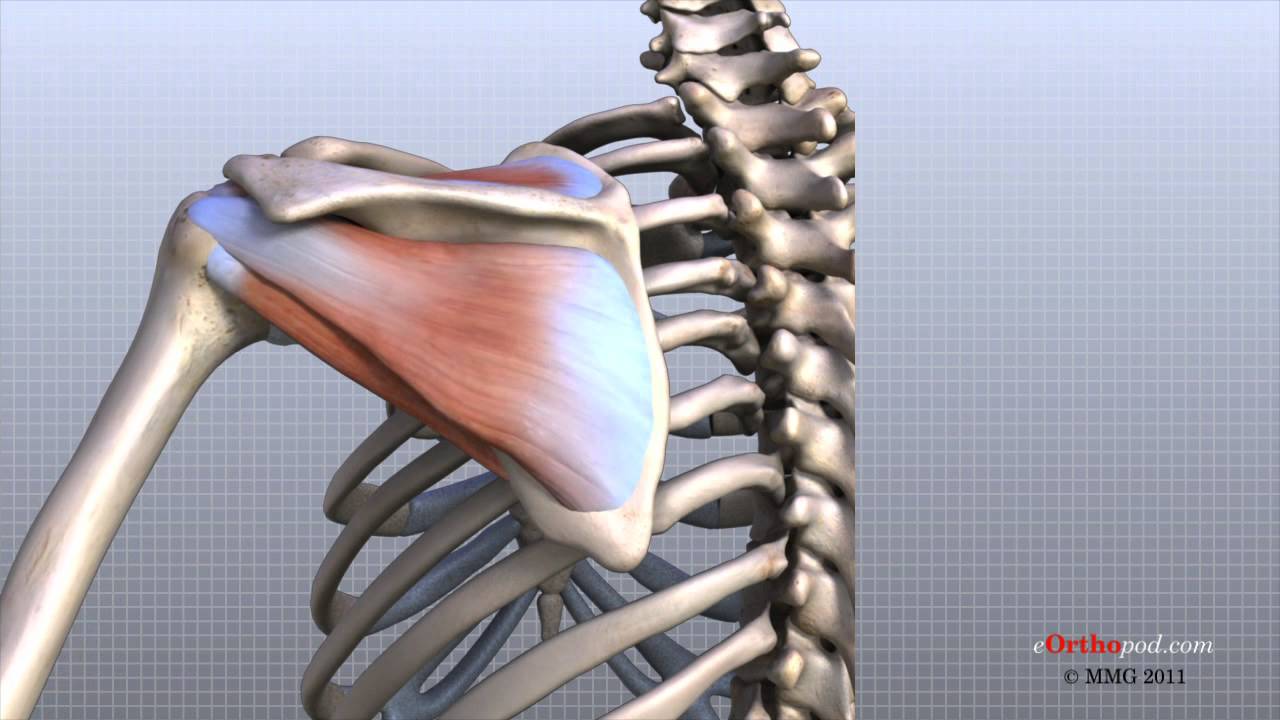
Right Shoulder Injuries High Impact Visual Litigation Strategies from res.cloudinary.com This diagram here just shows the joint capsule itself. Right shoulder joint arthrography coronal t1wi a coronal t2wi b download scientific diagram the diaphragm and liver in context. We'll remove the humerus and we'll take a look at the glenoid cavity. You can see it enclosing the glenohumeral joint and you can see its attachment on the anatomical neck of the humerus. We're looking laterally now at the right shoulders. Besides big lifting jobs, the shoulder joint is also responsible for getting the hand in the right position for any function. I sustained fractures to the right shoulder & top of arm in 2003. In this episode of eorthopodtv, orthopaedic surgeon randale c.
Explore more like shoulder joint anatomy diagram.
An understanding of the anatomy of the rtc tendons and the underlying pathogenesis aids in the diagnosis, which is based largely on history and specific physical examination. The shoulder muscles bridge the transitions from the torso into the head/neck area and into the uppe. Posted on december 13, 2018december 12, 2018. I sustained fractures to the right shoulder & top of arm in 2003. Movements of the human shoulder represent the result of a complex dynamic interplay of structural bony anatomy and biomechanics, static ligamentous and tendinous. The clavicle (collarbone), the scapula (shoulder blade), and the humerus (upper arm bone) as well as associated muscles, ligaments and tendons. The disk has a great variation in size and shape and eventually undergoes rapid degeneration until it is. Human anatomy diagrams show internal organs, cells, systems, conditions, symptoms and sickness information. We'll remove the humerus and we'll take a look at the glenoid cavity. When you realize all the different ways and positions we use our hands. Sechrest, md narrates an animated tutorial on the basic anatomy of the shoulder. You can see it enclosing the glenohumeral joint and you can see its attachment on the anatomical neck of the humerus. Anatomical diagram of the muscles of the neck.
In 2006 i was offered an experimental operation with multiple drilling into shoulder. 2.1 bones of the shoulder girdle. This diagram depicts shoulder muscles anatomy diagram. Ac joint is a diathrodial joint with a fibrocartilaginous disk. Anatomical diagram of the muscles of the neck.
Shoulder Anatomy Animated Tutorial Youtube from i.ytimg.com Anatomical diagram of the muscles of the neck. Blank head and neck muscles diagram | body muscles … from i.pinimg.com. Elbow dislocations constitute 10% to 25% of all injuries to the elbow. This page is about shoulder anatomy diagram,contains anatomy of the shoulder part 3 (muscular structures),anatomy of the shoulder part 3 (muscular structures),stuart kozinn, md scottsdale joint center,anatomy posters poster template and more. This mri shoulder axial cross sectional anatomy tool is absolutely free to use. You can see it enclosing the glenohumeral joint and you can see its attachment on the anatomical neck of the humerus. The shoulder joint is formed where the humerus (upper arm bone) fits into the scapula. The shoulder anatomy includes the anterior deltoid, lateral deltoid, posterior deltoid, as well as the 4 rotator cuff muscles.
When you realize all the different ways and positions we use our hands.
Explore more like shoulder joint anatomy diagram. Sechrest, md narrates an animated tutorial on the basic anatomy of the shoulder. The scapula (shoulder blade), clavicle (collarbone) and humerus. Hi, good explanation right there! Enjoy the videos and music you love, upload original content, and share it all with friends, family, and the world on youtube. Ap x ray of a dislocated right elbow. Wiring diagram for genie garage door opener. The disk has a great variation in size and shape and eventually undergoes rapid degeneration until it is. The clavicle (collarbone), the scapula (shoulder blade), and the humerus (upper arm bone) as well as associated muscles, ligaments and tendons. Human anatomy diagrams show internal organs, cells, systems, conditions, symptoms and sickness information. Shoulder joint anatomy shoulder joint muscles upper limb anatomy gross anatomy yoga anatomy scapula medical anatomy human anatomy and physiology muscle anatomy. Learn vocabulary, terms and more with flashcards, games and other study tools. Elbow dislocations constitute 10% to 25% of all injuries to the elbow.
Radiology department of the rijnland hospital, leiderdorp and the introduction shoulder anatomy diagram. Explore more like shoulder joint anatomy diagram.